Microfluidics is a technique to investigate highly efficiently chemical reactions in the liquid phase, conducting various chemical manipulations, such as phase separation, solvent extraction using a laminar flow, etc. In this study, the XAS measurements in the soft X-ray regions were applied to the element-selective analyses of liquid mixtures in a microfluidic flow [1]. Figure 1 shows the schematics of the XAS measurement system for microfluidics. The microfluidic cell consists of the T-shaped microfluidics with the width and depth of 50 μm made in PDMS resin and covered by the Si3N4 membrane with a thickness of 100 nm. The microfluidic cell was in an atmospheric helium condition, which was separated by the SiC membrane with the window size of 30 × 30 μm2 from the beamline under the ultrahigh vacuum condition. XAS spectra was measured by detecting fluorescent soft X-rays using a silicon drift detector.
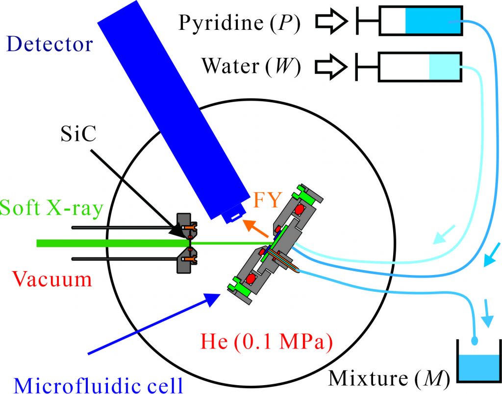
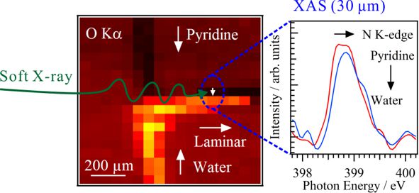
Figure 2 shows the soft X-ray fluorescent image of a T-shaped microfluidics in the O Kα regime by soft X-rays at 550 eV. Water and pyridine were merged at the center position, and a laminar flow of pyridine and water was clearly observed in the mixed part. The N K-edge XAS spectra of pyridine were measured in the mixed part with the spatial resolution of 30 × 30 μm2, which shows characteristic energy shifts near the liquid-liquid interface in a laminar flow. The distributions of the molar fractions of pyridine and water near the liquid-liquid interface have been determined from the energy shifts probed at different geometric positions, where pyridine is mixed in the water part of the laminar flow and vice versa. The present work clearly shows that the spatially-resolved XAS technique will be applicable for investigating the mechanisms of chemical and biological reactions prepared by microfluidics.