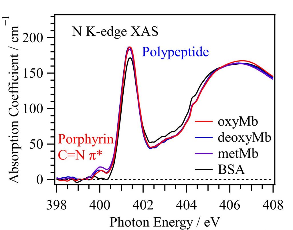
Spin states of myoglobin, which is one of hemoprotein, in aqueous solutions at room temperature were probed from the porphyrin C=N π* peaks of heme using the N K-edge XAS [1]. Figure 1 shows the N K-edge XAS spectra of myoglobin in different forms. Oxymyoglobin (oxyMb) contains a six-coordinated heme with a Fe2+ ion connected to an oxygen molecule. Deoxymyoglobin (deoxyMb) contains a five-coordinated heme with a Fe2+ ion. Metmyoglobin (metMb) contains a six-coordinated heme with a Fe3+ ion connected to a water molecule. The protein polypeptide chains contain numerous nitrogen atoms and show a strong peak at 401.4 eV. The XAS spectrum of bovine serum albumin (BSA) shows no peaks in the energy region of the C=N π* peaks because BSA does not have a heme. In the XAS spectra of oxyMb, deoxyMb, and metMb, the porphyrin C=N π* peaks of heme exist approximately at 400.0 eV and can be distinguished from the protein polypeptide peaks.
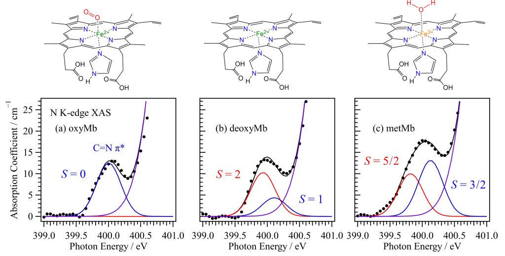
As shown in Fig. 2, the spin states of myoglobin heme irons were analyzed from the porphyrin C=N π* peaks compared with the inner-shell calculations. The XAS spectrum of oxyMb exhibits a single C=N π* peak and exclusively shows the S = 0 state. By contrast, the XAS spectra of deoxyMb and metMb exhibit two C=N π* peaks. DeoxyMb shows a spin equilibrium between the S = 2 and 1 states. Because of the larger intensity of the first C=N π* peak compared to the second peak, deoxyMb in the S = 2 state is favored over the S = 1 state. MetMb shows a spin equilibrium between the S = 5/2 and 3/2 states. Because the intensity of the second C=N π* peak is larger than that of the first peak, metMb in the S = 3/2 state is favored over the S = 5/2 state. Because the porphyrin C=N π* peaks reflect the electronic structures and spin multiplicities of heme due to metal‒ligand delocalization, the N K-edge XAS is suitable to investigate the spin equilibriums of hemoproteins in the physiological conditions, which are influenced by factors such as liquid temperature, solvent, gas adsorption, and protein structure.